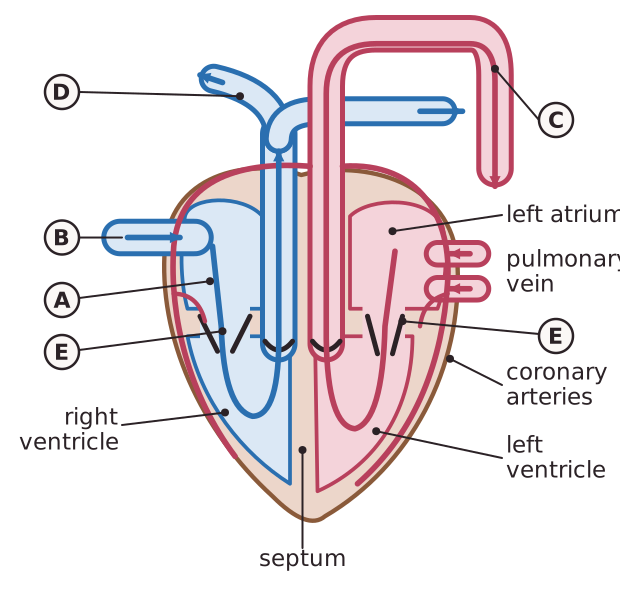
The mammalian heart is a muscular pump made of cardiac muscle, which contracts and relaxes without tiring. The heart muscle has its own blood supply, the coronary arteries, which bring it oxygen and glucose. The heart has four chambers. The two upper chambers, the atria, receive blood. The two lower chambers, the ventricles, pump it out. A wall called the septum keeps oxygenated and deoxygenated blood apart.
Blood from the body enters the right atrium through the vena cava, passes to the right ventricle, and leaves through the pulmonary artery to the lungs. Blood returns from the lungs in the pulmonary vein to the left atrium, passes to the left ventricle, and leaves through the aorta to the body. Valves keep blood flowing in one direction and stop it flowing backwards. The atrioventricular valves (the bicuspid valve on the left and the tricuspid valve on the right) lie between the atria and ventricles, and the semilunar valves lie at the exits to the pulmonary artery and aorta. The left ventricle has a thicker wall than the right because it pumps blood at higher pressure around the body, while the right ventricle pumps only to the lungs.
Arteries carry blood away from the heart at high pressure. They have thick walls containing muscle and elastic fibres, which stretch and recoil as the heart beats, and a narrow lumen. Veins carry blood back to the heart at low pressure. They have thinner walls, a wide lumen and valves that stop backflow. Capillaries have walls only one cell thick, so the diffusion distance is short. Blood moves slowly through them, not because they are narrow, but because their total cross-sectional area is far larger than that of the aorta. The slow flow gives time for exchange by diffusion.