Skip to content

Structure of the Liver

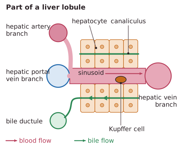
The liver is divided into lobes, which divide further into cylindrical lobules. Liver cells are called hepatocytes, have large nuclei and lots of Golgi and mitochondria. Blood from branches of the HPV and hepatic artery are mixed in spaces called sinusoids supplying cells with enough oxygen for their metabolic activity. Kupffer cells in the sinusoids are macrophages that engulf microorganisms and foreign particles and old red blood cells. The blood drains into a branch of the hepatic vein. Hepatocytes secrete bile into canaliculi. This drains into a branch of the bile duct (ductules) which leads to the gall bladder.
Read the text

Read the text and highlight anything you think is important. When you go on, the text is hidden and you answer from memory.

Specification: OCR Biology A (H420), A-level, 5.1.2 Excretion as an example of homeostatic control. Topic: Homeostasis.

Key terms in this lesson

Neurone
A specialised nerve cell adapted to transmit electrical impulses (nerve impulses) around the body.
Generator potential
A change in potential produced in a sensory receptor cell in response to a stimulus, which, if large enough, triggers an action potential.
Positive feedback
A control mechanism in which a change in a factor triggers a response that increases the change further, rather than reversing it.

More in Homeostasis

All 33 lessons in Homeostasis · All OCR A-level Biology topics