After gel electrophoresis the DNA bands cannot be seen, and the soft gel is hard to handle. Southern blotting transfers the DNA from the gel onto a membrane so specific fragments can be located with gene probes.Method
1. The gel is soaked in an alkaline solution. This denatures the DNA, separating it into single strands so a probe can bind later.
2. A nylon membrane is placed on the gel, with layers of absorbent paper on top. Solution is drawn up through the gel by capillary action, carrying the DNA fragments onto the membrane. The fragments keep the same positions they had in the gel, so the membrane is an exact copy of the pattern.
3. The DNA is fixed to the membrane, using ultraviolet light or heat.
4. The membrane is placed in a solution containing the gene probe.
5. The membrane is washed to remove any probe that has not bound.
6. The position of the probe is detected.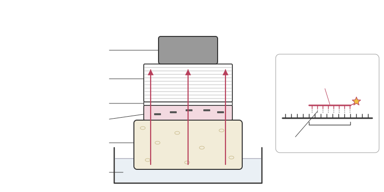
A gene probe is a short, single-stranded piece of DNA with a base sequence complementary to the target sequence. It is labelled so that it can be detected, either with a radioactive isotope such as 32P or with a fluorescent tag. The probe binds (hybridises) to the target by complementary base pairing, and only to fragments carrying that sequence.A radioactive probe is located by placing X-ray film over the membrane: the film darkens where the probe has bound. This is called autoradiography. A fluorescent probe glows under ultraviolet light.The result is a pattern of bands showing only the fragments that contain the target sequence.
1. The gel is soaked in an alkaline solution. This denatures the DNA, separating it into single strands so a probe can bind later.
2. A nylon membrane is placed on the gel, with layers of absorbent paper on top. Solution is drawn up through the gel by capillary action, carrying the DNA fragments onto the membrane. The fragments keep the same positions they had in the gel, so the membrane is an exact copy of the pattern.
3. The DNA is fixed to the membrane, using ultraviolet light or heat.
4. The membrane is placed in a solution containing the gene probe.
5. The membrane is washed to remove any probe that has not bound.
6. The position of the probe is detected.
A gene probe is a short, single-stranded piece of DNA with a base sequence complementary to the target sequence. It is labelled so that it can be detected, either with a radioactive isotope such as 32P or with a fluorescent tag. The probe binds (hybridises) to the target by complementary base pairing, and only to fragments carrying that sequence.A radioactive probe is located by placing X-ray film over the membrane: the film darkens where the probe has bound. This is called autoradiography. A fluorescent probe glows under ultraviolet light.The result is a pattern of bands showing only the fragments that contain the target sequence.