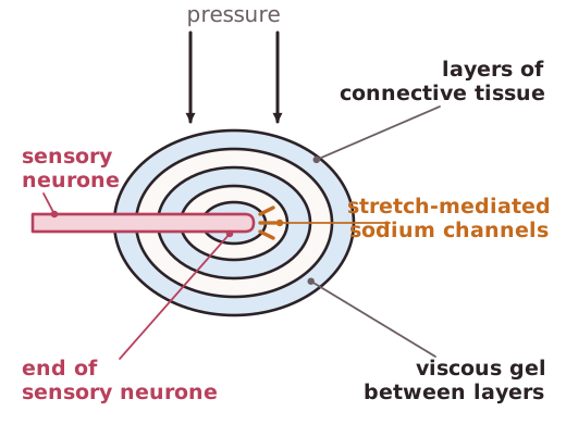
Pacinian corpuscles are located deep within the skin and detect mechanical pressure. Most are found in the fingers, soles of the feet and external genitalia but some are also found within the joints, ligaments and tendons so the organism can detect which joints are changing direction.The tip of a sensory neurone is in the middle of the Pacinian corpuscle which is made of layers of connective tissue separated by a viscous gel.The membrane at the tip of the sensory neurone can be stretched by pressure applied to the Pacinian corpuscle. This causes sodium channels called stretch-mediated sodium channels near the tip of the sensory neurone to be stretched. The sodium channels widen and allow the diffusion of sodium ions into the neurone.The entry of sodium ions into the neurone reverses the polarity of the membrane. The membrane is said to be depolarised.The potential difference generates the nerve impulse and is known as the generator potential.