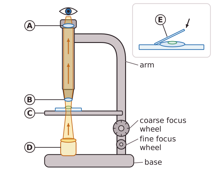
A light microscope uses a lamp to shine light up through a specimen on the stage. The specimen is placed on a glass slide, usually with a drop of water, and a thin cover slip is lowered on top. The cover slip holds the specimen flat and stops it drying out. Light passes through the specimen and then through the lenses. The eyepiece lens and an objective lens each magnify the image, and the total magnification is the eyepiece magnification multiplied by the objective magnification.
Many cells are almost colourless, so they are hard to see. A stain is a dye that colours the specimen. Different stains are taken up by different structures, so a stain can show up the nucleus or highlight different tissues. Iodine solution stains onion cells and methylene blue stains cheek cells, for example.
Magnification tells you how many times bigger the image is than the real object: magnification = image size ÷ real size. If the image size is measured in millimetres and the real size is in micrometres, convert first, because 1 mm = 1000 µm. A cell that is 50 µm long and appears 5 mm long in a drawing is magnified 100 times (5000 ÷ 50).