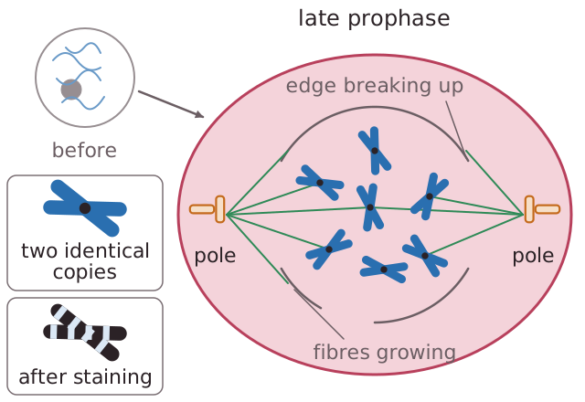
1. Chromatin fibres condense and supercoil to form chromosomes. The chromosomes become visible under a light microscope.2. Cylindrical bundles of proteins called the centrioles move to either end of the cell.3. The nucleolus disappears and the nuclear membrane disintegrates.4. Microtubules form the nuclear spindle from the centrioles, stretching from the poles to the equator.5. The spindle fibres attach to the centromeres and start moving the chromosomes to the middle of the cell.6. By the end of prophase the nuclear membrane has disappeared.
Read the text
Read the text and highlight anything you think is important. When you go on, the text is hidden and you answer from memory.
Specification: OCR Biology A (H020), AS-level, 2.1.6 Cell division, cell diversity and cellular organisation. Topic: Cell Division.
Key terms in this lesson
- nucleolus
- A spherical structure within the nucleus where ribosomal RNA is made and combined with protein to form ribosomes.
- centriole
- A structure made of microtubules that organises the spindle fibres during cell division.
- chromatin
- The complex of DNA loosely coiled around histone proteins found in the nucleus between cell divisions.
- centromere
- The region of a chromosome that joins two sister chromatids together and attaches to the spindle fibres.
More in Cell Division
All 31 lessons in Cell Division · All OCR AS-level Biology topics