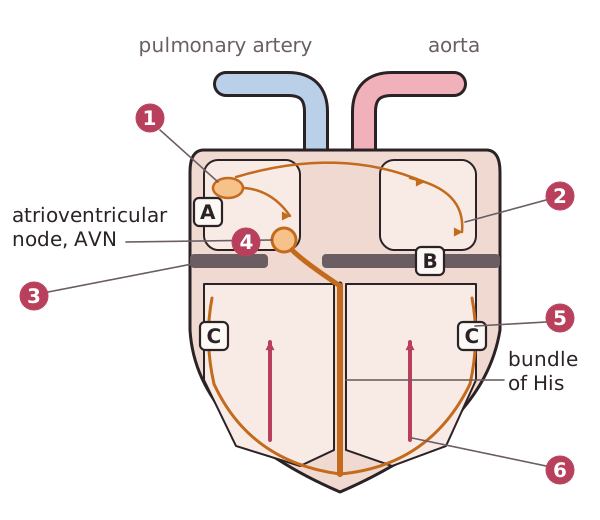
Cardiac muscle is myogenic, but the heartbeat still needs to be coordinated so that the atria contract before the ventricles. This is done by specialised tissue in the heart that generates and conducts waves of electrical excitation.1. Sinoatrial node (SAN). The SAN is a small patch of tissue in the wall of the right atrium. It acts as the pacemaker, generating a wave of electrical excitation at regular intervals.2. Spread across the atria. The wave spreads across the walls of both atria, causing them to contract (atrial systole).3. Non-conducting tissue. A layer of non-conducting tissue between the atria and the ventricles stops the wave passing directly into the ventricle walls.4. Atrioventricular node (AVN). The only route through is the AVN, which delays the impulse for about 0.1 s. This delay gives the atria time to finish contracting and the ventricles time to fill before they contract.5. Bundle of His and Purkyne fibres. The AVN passes the impulse down the bundle of His, conducting fibres in the septum, to the apex (bottom) of the heart. From there it spreads up through the walls of the ventricles along the Purkyne fibres.6. Ventricular systole. The ventricles contract from the apex upwards. This squeezes blood up and out of the ventricles into the arteries, which leave from the top of the ventricles, rather like squeezing a tube of toothpaste from the bottom.