 BRAINCAKE
PET Scanning
PET (positron emission tomography) uses a positron-emitting tracer, usually fluorine-18 in FDG, to make 3D images of active tissue such as tumours or regions of the brain.
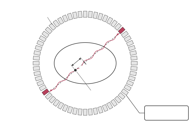
1. The tracer collects in the active tissue and emits positrons.
2. Each positron travels less than about a millimetre, meets an electron and they annihilate.
3. Their mass becomes two gamma photons of equal energy that travel in opposite directions. Each photon has energy equal to the rest energy of an electron:
Sign in free to see the rest of this lesson, the R.E.C.I.P.E. recall steps and the quiz
BrainCake is free. Make an account in seconds and pick up where this page stops.
Key terms in this lesson
- annihilation
- The process in which a particle and its antiparticle collide and their mass is completely converted into energy, usually as gamma photons.
More in Medical Imaging
- Medical Tracers
- Technetium-99m and Fluorine-18
- The Gamma Camera
- Diagnosis with a Gamma Camera
- Ultrasound
- The Piezoelectric Effect
- Ultrasound Transducers
- A-scans and B-scans
All 20 lessons in Medical Imaging · All OCR A-level Physics topics