A gamma camera detects the gamma photons emitted by a tracer inside the patient and forms an image of where the tracer is.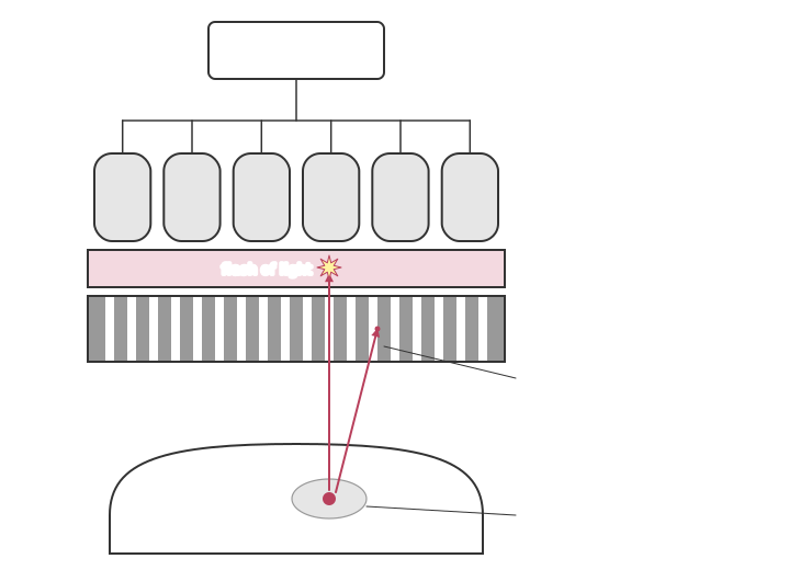
• Scintillator: a large crystal of sodium iodide. Each gamma photon that is absorbed produces thousands of visible light photons.
• Photomultiplier tubes: a hexagonal array of tubes behind the crystal. Each converts the light into an electrical pulse and amplifies it greatly.
• Computer and display: the tube nearest the flash gives the largest pulse, so the computer compares the sizes of the pulses from nearby tubes to locate where each gamma photon struck. Thousands of such events build up the image.The brightness at each point shows how much tracer is in the tissue below. The collimator throws away most gamma photons, so the scan takes several minutes to collect enough counts for a clear image.
• Scintillator: a large crystal of sodium iodide. Each gamma photon that is absorbed produces thousands of visible light photons.
• Photomultiplier tubes: a hexagonal array of tubes behind the crystal. Each converts the light into an electrical pulse and amplifies it greatly.
• Computer and display: the tube nearest the flash gives the largest pulse, so the computer compares the sizes of the pulses from nearby tubes to locate where each gamma photon struck. Thousands of such events build up the image.The brightness at each point shows how much tracer is in the tissue below. The collimator throws away most gamma photons, so the scan takes several minutes to collect enough counts for a clear image.