Ultrasound scans use the time taken for echoes to return from boundaries inside the body. A pulse travels to a boundary and back, so the depth d of the boundary isd = ct / 2 where c is the speed of ultrasound in the tissue and t is the time between sending the pulse and receiving its echo.A-scan (amplitude scan): a single transducer sends pulses along one line. The echoes are shown as peaks on a graph of amplitude (voltage) against time. The position of each peak gives the depth of a boundary, and its height shows how strongly that boundary reflects. A-scans are used to measure distances accurately, for example the depth of the eyeball or the thickness of tissue.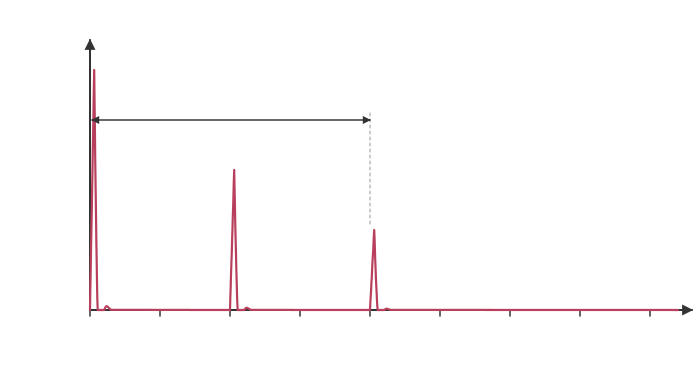
d = 1500 × 40 × 10−6 / 2 = 0.030 m = 30 mm B-scan (brightness scan): each echo is shown as a dot on a screen whose brightness depends on the strength of the echo, at a position given by the direction of the pulse and the echo time. The transducer is moved across the skin, or an array of transducers is used, so many lines are combined into a two-dimensional (2D) image of a slice through the body. B-scans are used for pregnancy scans and for imaging organs, and can be updated many times a second to give a moving image.