Doppler imaging uses ultrasound to measure the speed of blood flow, for example to find narrowed arteries or check blood flow through the heart and the placenta.Ultrasound of frequency f is directed at a blood vessel. It is reflected by moving red blood cells, and the reflected ultrasound has a changed frequency because of the Doppler effect. If the blood is moving towards the transducer the reflected frequency is higher; if it is moving away, the reflected frequency is lower.The change in frequency (Doppler shift) isΔf = 2fv cos θ / c where v is the speed of the blood, c the speed of ultrasound in tissue and θ the angle between the ultrasound beam and the direction of blood flow. The factor of 2 appears because the shift happens twice: the moving cells receive a shifted frequency, then reflect it as a moving source. If the beam is at 90° to the flow, cos θ = 0 and there is no shift, so the transducer is held at an angle to the vessel.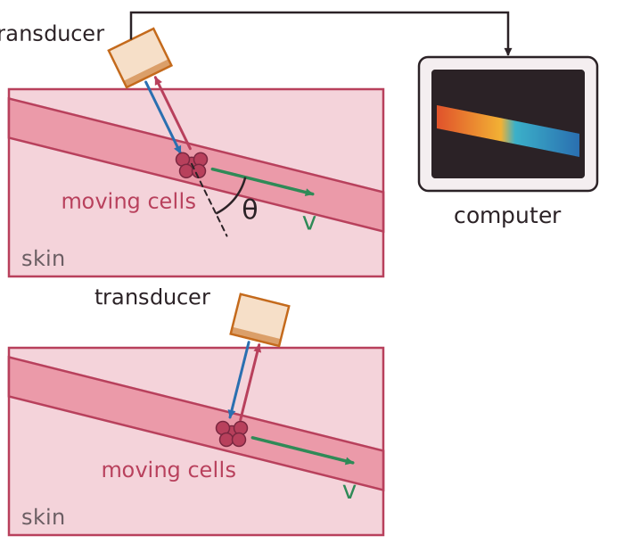 Worked example: f = 5.0 MHz, v = 0.30 m s−1, θ = 30°, c = 1500 m s−1.
Δf = 2 × 5.0 × 106 × 0.30 × cos 30° / 1500 = 1.7 × 10³ Hz The shift of 1.7 kHz is in the audible range, so the machine can also play it as a sound. The computer converts Δf into a speed and shows the flow in colour on the image.
Worked example: f = 5.0 MHz, v = 0.30 m s−1, θ = 30°, c = 1500 m s−1.