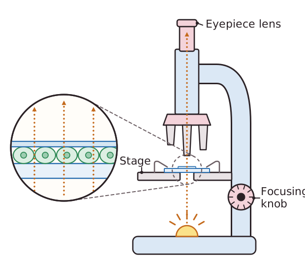
A light microscope uses light and lenses to make a small object look bigger. Light from the lamp passes up through a thin specimen resting on the stage. The specimen is placed on a glass slide, and a cover slip is lowered gently on top. The cover slip holds the specimen flat and protects the objective lens. Only thin specimens work, because light has to pass through them.
Most cells are colourless, so they are hard to see. A stain is a dye that colours the cells, or colours different structures and tissues so that they stand out. Methylene blue is often used on cheek cells and iodine solution on onion epidermis cells.
The light passes through two sets of lenses. The eyepiece lens and the objective lens each magnify the image. You find the total magnification by multiplying the eyepiece magnification by the objective magnification, so a ×10 eyepiece with a ×10 objective gives ×100. For a drawing or a photograph, magnification = image size ÷ actual size, using the same units for both.