 BRAINCAKE
Ultrastructure
The ultrastructure of a cell is the detailed internal structure that can only be seen with an electron microscope. A light microscope shows the nucleus and cytoplasm; the electron microscope reveals the membranes and organelles inside.
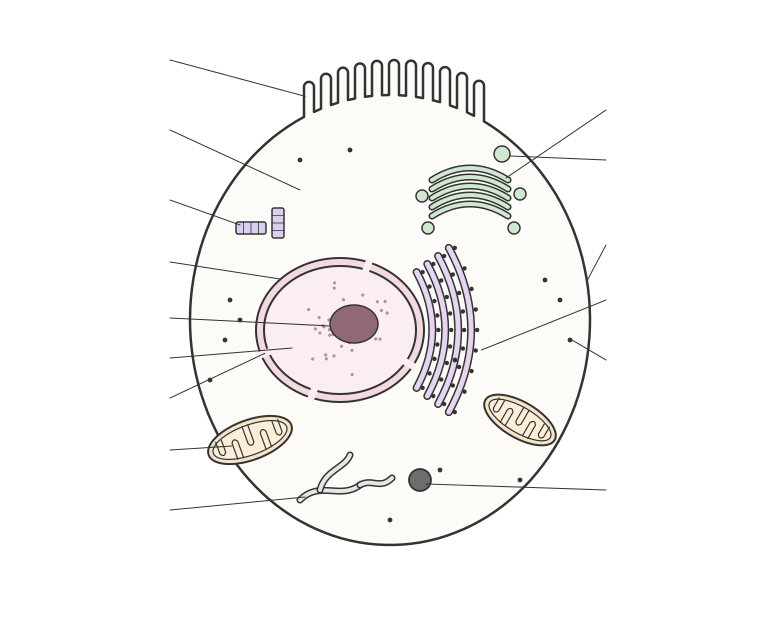
An electron micrograph of an animal cell (a eukaryotic cell) typically shows:
• nucleus with a double nuclear envelope, nuclear pores and a darker nucleolus
• rough endoplasmic reticulum (flattened sacs lined with ribosomes) and smooth endoplasmic reticulum (tubes without ribosomes)
• Golgi apparatus, a stack of curved flattened sacs with vesicles
• mitochondria, with a double membrane and folded cristae
Sign in free to see the rest of this lesson, the R.E.C.I.P.E. recall steps and the quiz
BrainCake is free. Make an account in seconds and pick up where this page stops.
Key terms in this lesson
- nuclear envelope
- The double membrane that surrounds the nucleus, perforated by nuclear pores.
More in Eukaryotic Cells
- Light Microscope
- Electron Microscope
- Transmission Electron Microscope
- Scanning Electron Microscope
- Membranes
- Phospholipid Bilayer
- Membrane Proteins
- Fluid Mosaic Model
All 30 lessons in Eukaryotic Cells · All Edexcel AS-level Biology topics