Skip to content

Light Microscope

The light microscope (optical microscope) uses visible light focused by glass lenses. Light passes up through a thin specimen on a slide, then through the objective lens and the eyepiece lens. The total magnification is the product of the two:
total magnification = objective × eyepiece
so a ×40 objective with a ×10 eyepiece gives ×400.
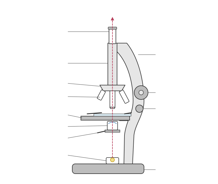
Because the wavelength of light is relatively long, the resolution is limited to about 0.2 µm (200 nm) and the useful magnification to about ×1500. You can see cells, nuclei and, with care, chloroplasts and mitochondria as dots, but not the detailed structure of organelles or ribosomes.Specimens are usually thin sections, often stained (for example iodine for starch, methylene blue for animal cells) to add contrast. A big advantage is that living cells can be viewed, in colour, and the equipment is cheap and portable.To measure cells, an eyepiece graticule (a scale in the eyepiece) is calibrated against a stage micrometer, a slide with a scale of known divisions, often 0.01 mm (10 µm) apart. For example, if 40 graticule divisions line up with 10 stage divisions (100 µm), one graticule division = 100 ÷ 40 = 2.5 µm at that objective. The calibration must be repeated for each objective lens.

Read the text

Read the text and highlight anything you think is important. When you go on, the text is hidden and you answer from memory.

Specification: Edexcel Biology B (8BI0), AS-level, 2.1 Eukaryotic and prokaryotic cell structure and function. Topic: Eukaryotic Cells.

Key terms in this lesson

resolution
The smallest distance between two points that can still be seen as two separate points; higher resolution shows more detail.

More in Eukaryotic Cells

All 30 lessons in Eukaryotic Cells · All Edexcel AS-level Biology topics