The eye is a sense organ that focuses light onto a layer of photoreceptors, the retina, and sends information to the brain along the optic nerve.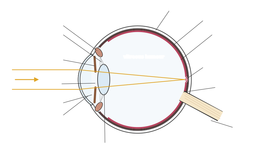
• Cornea: the transparent front part of the outer coat. It does most of the refraction (bending) of light entering the eye.
• Iris: the coloured ring of muscle that controls the size of the pupil, the hole through which light enters. In bright light the pupil constricts; in dim light it dilates. This is a reflex controlled by the autonomic nervous system.
• Lens: a transparent, flexible structure that makes fine adjustments to focus. Its shape is changed by the ciliary muscles acting through the suspensory ligaments.
• Aqueous humour and vitreous humour: transparent fluids that keep the eye's shape and let light through.
• Choroid: a dark, pigmented layer containing blood vessels that absorbs stray light and supplies the retina.
• Retina: the inner layer containing the photoreceptors, rods and cones.
• Fovea: a small area of the retina with the highest density of cones, giving the sharpest vision.
• Blind spot: where the optic nerve leaves the eye. There are no photoreceptors here, so light falling on it is not detected.
• Optic nerve: carries impulses from the retina to the brain.So light passes through the cornea, aqueous humour, pupil, lens and vitreous humour before it reaches the retina.
• Cornea: the transparent front part of the outer coat. It does most of the refraction (bending) of light entering the eye.
• Iris: the coloured ring of muscle that controls the size of the pupil, the hole through which light enters. In bright light the pupil constricts; in dim light it dilates. This is a reflex controlled by the autonomic nervous system.
• Lens: a transparent, flexible structure that makes fine adjustments to focus. Its shape is changed by the ciliary muscles acting through the suspensory ligaments.
• Aqueous humour and vitreous humour: transparent fluids that keep the eye's shape and let light through.
• Choroid: a dark, pigmented layer containing blood vessels that absorbs stray light and supplies the retina.
• Retina: the inner layer containing the photoreceptors, rods and cones.
• Fovea: a small area of the retina with the highest density of cones, giving the sharpest vision.
• Blind spot: where the optic nerve leaves the eye. There are no photoreceptors here, so light falling on it is not detected.
• Optic nerve: carries impulses from the retina to the brain.So light passes through the cornea, aqueous humour, pupil, lens and vitreous humour before it reaches the retina.