 BRAINCAKE
Structure and Function of the Kidney
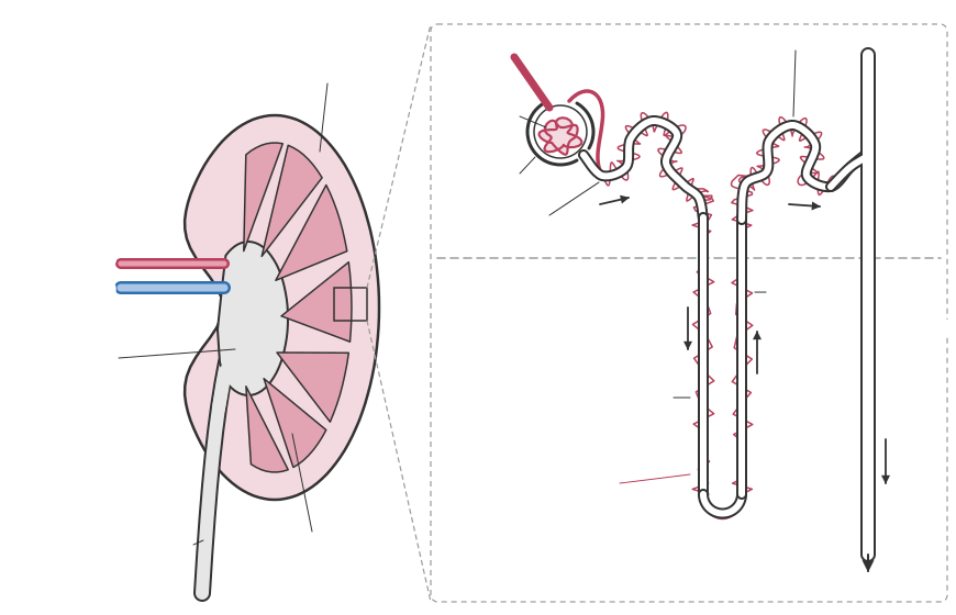
Humans have two kidneys at the back of the abdomen. Each receives blood from a renal artery, a branch of the aorta, and returns it through a renal vein to the vena cava. Urine leaves each kidney through a ureter to the bladder, and is released through the urethra.
A kidney cut lengthways shows three regions:
• the outer cortex, containing the Bowman's capsules and convoluted tubules
• the inner medulla, containing the loops of Henle and collecting ducts, arranged in cone-shaped pyramids
• the renal pelvis, a cavity that collects urine and leads into the ureter
Sign in free to see the rest of this lesson, the R.E.C.I.P.E. recall steps and the quiz
BrainCake is free. Make an account in seconds and pick up where this page stops.
More in Homeostasis
- Additional Responses
- Protein Homeostasis in the Liver
- Osmoregulation
- Osmoregulation in Mammals
- Ultrafiltration
- Selective Reabsorption
- Proximal Convoluted Tubule
- Loop of Henle
All 41 lessons in Homeostasis · All Edexcel A-level Biology topics