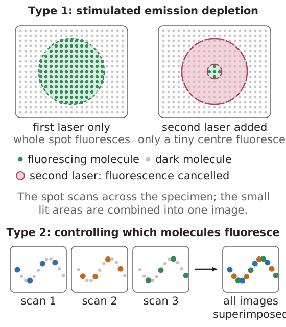
Super resolved fluorescence microscopy can achieve a resolution of about 0.02μm (20nm) using visible light.There are two different types. One combines many very small scanned areas to produce a sharper image and the other superimposes many images with normal resolution to produce a high resolution image.The first type uses the technique of stimulated emission depletion.Two lasers are directed onto a specimen. The first scans the specimen and causes fluorescence and the second slightly offset laser cancels the fluorescence from all but a small molecular-sized area of the specimen.The second process involves controlling which molecules fluoresce. Different molecules are allowed to fluoresce each time the specimen is scanned.The images are superimposed to produce a high resolution image.The path of individual molecules can be traced during cellular processes.