In atomic force microscopy, the surface of the specimen is scanned with a mechanical probe with a sharp tip to generate a 3D image of the specimen.
The probe has a sharp tip and is supported by a cantilever.When the tip is brought close to the specimen the force between the tip and specimen moves the cantilever. A laser beam is directed onto the cantilever and reflects off the cantilever onto the detector.Changes in the intensity of the reflected light due to the movement of the cantilever cause an image to be formed.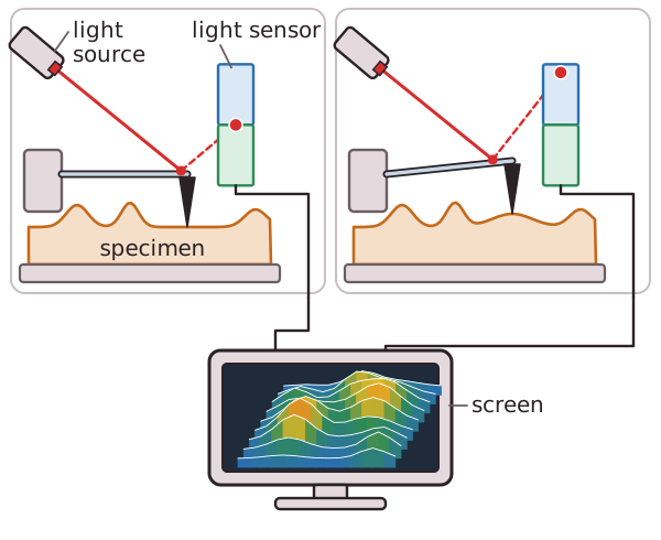 Fixation and staining are not required.The resolution is as high as 0.1nm and living systems can be examined without damaging them.
The probe has a sharp tip and is supported by a cantilever.When the tip is brought close to the specimen the force between the tip and specimen moves the cantilever. A laser beam is directed onto the cantilever and reflects off the cantilever onto the detector.Changes in the intensity of the reflected light due to the movement of the cantilever cause an image to be formed.
Fixation and staining are not required.The resolution is as high as 0.1nm and living systems can be examined without damaging them.