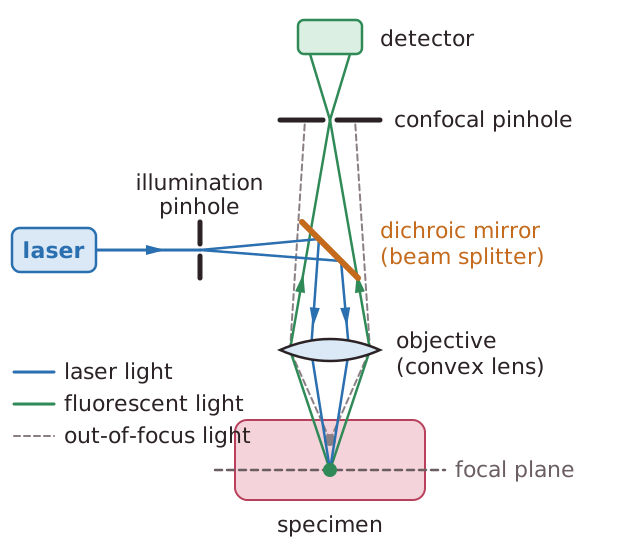
A laser is a very intense beam of light.A laser scanning confocal microscope uses point illumination.Selective components are labelled with a fluorescent dye.A single spot of focused laser light is moved across the specimen and the components which are labelled emit fluorescent light.Only light close to the focal plane is detected so it gives a very sharp two-dimensional image. Light from different focal planes would reduce the resolution and cause blurring.Light passing through the illumination pinhole is reflected off a dichroic mirror (beam splitter) onto the sample.The Light reflecting off the sample is focused by a convex lens so that it passes through a confocal pinhole to the detector.The confocal pinhole is the same distance from the mirror as the illuminating pinhole so fluorescent light and light from the laser travel the same path and have the same focal plane.The technique is non-invasive and can also be used to diagnose diseases of the eye.