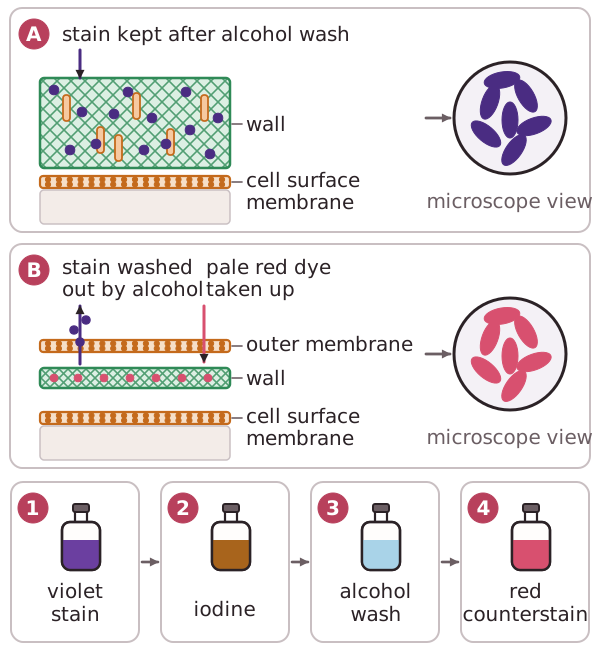
All bacterial cell walls are made of peptidoglycan. Before staining bacteria are often colourless. Gram positive bacteria have a thick net-like layer of peptidoglycan containing a substance called teichoic acid.When crystal violet followed by iodine is applied, the crystal violet-iodine complex is trapped within the cell wall and is not washed away when the sample is dehydrated with alcohol.When the cells are counterstained with safranin dye they do not pick up the red stain and appear blue or purple under the microscope.Gram negative bacteria have cell walls made from a thin layer of peptidoglycan with no teichoic acid and an outer lipopolysaccharide membrane.When dehydrated with alcohol after the Gram stain is applied, they do not retain the crystal-violet-iodine complex so will take up the red safranin stain when applied and appear red under the microscope.